Q. Your previous post about MRI’s brought up a question I have always wondered. When do you need an MRI versus a CAT Scan versus an X-ray? How do you know which type of imaging you need? -Jeanne
A. Thanks, Jeanne, for a great follow up question from Q & A: Do I Need an MRI for Low Back Pain? Many different types of imaging, such as an X-ray, CT Scan, and MRI, can be used depending on the situation and the structure being assessed. Let’s go through a quick synopsis of the basics on imaging. In addition, I will review the differences between a bone scan and a bone density scan, which is very important in diagnosing osteoporosis.
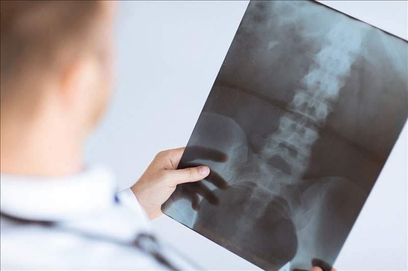
An X-ray, also known as radiography or a Roentgen ray, is an electromagnetic wave. The images produced show the parts of your body in different shades of black and white. This is because different tissues absorb different amounts of radiation (X-ray). The more radiation absorbed, the whiter it appears on the film. For example, calcium in bones absorbs X-rays the most, so bones appear white. Fat and other soft tissues absorb less and appear gray. Air absorbs the least, so the lungs appear black. If you have any metal implants, they will appear the most white.
X-rays are typically used to examine bones for a fracture or injury. Chest X-rays are often used to quickly spot pneumonia. Mammograms use X-rays to search for breast cancer. Fluoroscopy is a type of live action X-ray in which the results are delivered in real time. This is commonly used during certain types of spinal injections.
A computed tomography CT Scan (also known as a CAT Scan) uses X-rays to produce detailed images of the structures of the body. The CT scanner sends the X-rays through the body part/area being studied by taking very thin slices (images) of the targeted organ or area. They are then grouped back together by a computer to make a comprehensive image. CT Scans are best suited for viewing bone injuries, diagnosing lung and chest problems, and detecting cancer. CT Scans are widely used in Emergency Departments (ED’s) because the scan takes fewer than 5 minutes to perform depending on location of the scan. CT Scans produce more radiation than a typical X-ray or an MRI.
An MRI (Magnetic Resonance Imaging) is best suited for examining soft tissues, such as ligaments and tendons, spinal cord injuries, and brain tumors. Unlike an X-ray or a CT Scan, a MRI can take 30 minutes or more depending on the area and the detail of the scan. Like a CT Scan, the MRI takes images in slices, and then a computer recreates the information to give a comprehensive image. The smaller the slices taken, the more detailed and accurate the image meaning that the scan will take longer and cost more. An MRI typically costs more than a CT Scan. One advantage of an MRI is that it doesn’t use radiation like a CAT Scan.
A bone scan is a nuclear imaging test for looking at specific bone related injuries or disease. One advantage of a bone scan is that it can often discover a problem days to months earlier than a regular X-ray test.
During the scan, a radioactive substance called a “tracer” is injected into a vein in your arm. The tracer travels through your bloodstream and into your bones. Then a special camera takes images of the tracer in your bones. Areas that absorb little or no amount of tracer appear as dark or “cold” spots. This could show a lack of blood supply to the bone or certain types of cancer.
Areas of fast bone growth or repair absorb more tracer and show up as bright or “hot” spots in the images. Hot spots may point to problems such as arthritis, a tumor, a fracture or an infection. The level of detail is not always as good for a bone scan. However, bone scans can be very helpful for your physician in order to gain a proper diagnosis if you are experiencing unexplainable skeletal pain, bone infection or a bone injury that can’t be seen on a standard X-ray.
A bone density scan, also known as a Dual-energy X-ray absorptiometry (DXA, previously DEXA), is not the same as a bone scan. This particular scan measures the density of the bones by using two X-ray beams, each with different energy levels. One beam is high energy while the other is low energy. The amount of X-rays that pass through the bone is measured for each beam. This will vary depending on the thickness of the bone. Based on the difference between the two beams, the bone density can be measured.
The actual radiation exposure is low and typically less than a standard X-ray. A bone density scan is not only quick and safe, but it’s very important in diagnosing osteoporosis, which is the thinning of bones to the point they can become brittle and break. The National Osteoporosis Foundation recommends bone density scans for:
- Women who are 65 years and older
- Men who are 70 years and older
- Those who have broken a bone after the age of 50
- Women of menopausal age with risk factors for osteoporosis
- Post-menopausal women under the age of 65 with risk factors for osteoporosis
- Men between 50-69 years old with risk factors for osteoporosis
Thanks, Jeanne, for the question! The only way you can insure that you are receiving the best possible care for you and your loved ones is to understand more about your body and the medical options available today. Then you can research certain topics more thoroughly and have a complete, straight forward dialog with your medical providers.
I strongly believe that it is critical for all individuals to increase his/her knowledge base on basic medicine, health, fitness, and nutrition. My ultimate goal for The Physical Therapy Advisor is to help you in providing that education. What are your pains? What questions do you have? Please submit them to contact@thePhysicalTherapyAdvisor.com. I look forward to providing you with useful and practical types of “how to” information and to answer your health related questions. You can achieve optimal health!
Don’t forget subscribe to my e-mail newsletter! I will send you weekly posts on how to maximize your health, self-treat those annoying orthopaedic injuries, and gracefully age. To thank you for subscribing, you will automatically gain access to my FREE resource, My Top 8 Stretches to Eliminate Neck, Upper Back, and Shoulder Pain.
Be sure to join our growing community on Facebook by liking The Physical Therapy Advisor where you will receive additional health and lifestyle information!

